Dr Manish Singhal - The best Cancer Specialist in Delhi
Gall Bladder Cancer Specialist in Noida, Delhi NCR
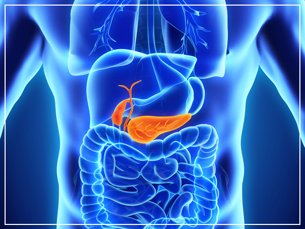
Gall bladder Cancer
Gallbladder cancer is a kind of disease that begins in the gallbladder. A gallbladder is a little, pear-formed organ under the liver. Both the liver and the gallbladder are the behind the lower ribs. In grown-ups, the gallbladder is more often than not around 3 to 4 inches long and ordinarily no more extensive than an inch. It concentrates and stores bile, a liquid made in the liver. Bile helps process the fats in nourishment as they go through the small digestive tract. Bile is either discharged from the liver specifically into pipes that convey it to the small digestive tract or is put away in the gallbladder and discharged later. Whenever sustenance (particularly greasy nourishment) is being processed, the gallbladder contracts and discharges bile through a little tube called the cystic pipe. The cystic channel cooperates with the basic hepatic conduit, which originates from the liver, to shape the regular bile pipe. The basic bile pipe joins the fundamental conduit from the pancreas (the pancreatic channel) to purge into the duodenum (the initial segment of the small digestive tract) at the ampulla of Vater. The gallbladder is useful, however, you needn’t bother with it to live. Numerous individuals have their gallbladders evacuated and go ahead to live normal lives.
The 5-year survival rate of gallbladder cancer is 19%, but it depends on several factors including but not limited to, the type of gallbladder cancer, the location of cancer, and the stage. Treatment is available in almost all situations. But the Covid-19 pandemic has not only caused increased fear among the immunocompromised cancer patients but, it has also made the maintenance of such treatment difficult. Dr. Manish Singhal, the best Oncologist in Noida, and his team are battling this difficulty successfully by offering chemotherapy at home, video consultations, online check-ins, and other services while taking preventive measures abiding by Covid-19 regulations.
Types of Gall bladder Cancers
There are lots of various sorts of cells in the gallbladder. Any of these cells composes could, in theory, form into a disease that is the reason there is more than one sort of gallbladder More than 85 out of each 100 gallbladder cancers (85%) are adenocarcinomas. This implies disease began in organ cells in the gallbladder lining. The organ cells regularly deliver bodily fluid (thick liquid). There are three sorts of adenocarcinomas of the gallbladder.
- Non-papillary adenocarcinoma
- Papillary adenocarcinoma
- Mucinous adenocarcinoma
More than 75 out of each 100 gallbladder malignancies (75%) are non-papillary adenocarcinomas. Just around 6 out of each 100 analyzed gallbladder cancers (6%) are papillary adenocarcinomas. These create in the tissues that hold the gallbladder set up (connective tissues). This sort of gallbladder cancer is less inclined to spread to the liver and close-by lymph hubs. It has a tendency to have a superior standpoint than most different sorts of gallbladder disease. With mucinous adenocarcinomas, the tumor cells are regularly in pools of bodily fluid, which is the means by which malignancy gets its name. Just around 1 or 2 out of each 100 gallbladder diseases (1 or 2%) are mucinous adenocarcinomas.
Squamous cell cancer of the gallbladder
Squamous cell cancers develop from the skin like cells that shape the coverage of the gallbladder, alongside the organ cells. Specialists treat these diseases similarly as adenocarcinomas.
Adenosquamous cancer of the gallbladder
Adenosquamous carcinomas are cancers that have both squamous cancer cells and glandular tumor cells. Your specialist may call this a blended histology. Specialists treat these tumors similarly as adenocarcinomas.
Small cell cancer of the gallbladder
Small cell carcinomas are additionally called oat cell carcinomas. They are called this in light of the fact that the tumor cells are an unmistakable oat shape.
Sarcoma of the gallbladder
Sarcoma is the name for cancer that influences the steady or ensuring tissues of the body – likewise called the connective tissues. Muscles, veins, and nerves are on the whole connective tissues. Cancer that starts in the muscle layer of the gallbladder is a sarcoma.
Neuroendocrine Cancer of the gallbladder
Neuroendocrine cancers are uncommon cancers that develop from hormone-delivering tissues, more often than not in the stomach related system. The most well-known sort of neuroendocrine tumor is called carcinoid.
Symptoms
According to our seasoned cancer doctor in delhi, Gallbladder cancer does not for the most part cause signs or side effects until over the span of the illness, however here and their indications can show up sooner and prompt an early determination. On the off chance that the cancer is found at an early stage, then treatment may be more powerful.
Some of the more common symptoms of gallbladder cancer are:
Stomach Pain
Many people with gallbladder cancer will have stomach pain. Mostly. in the upper portion of the stomach.
Vomiting
Individuals with gallbladder cancer in some cases have ‘vomiting’ as a side effect.
Lumps in the belly
If the tumor hinders the bile pipes, the gallbladder can swell to bigger than typical. Gallbladder disease can likewise spread to close-by parts of the liver. These can some of the time be felt by the specialist as lumps on the correct side of the stomach. They can likewise be distinguished by imaging tests, for example, an ultrasound.
Other common symptoms are:
- Loss of appetite
- Weight reduction
- Swelling in the belly
- Fever
- Irritated skin
- Dark urine
Gallbladder tumor isn’t normal, and these indications and signs will probably be caused by an option that is other than gallbladder cancer. For instance, individuals with gallstones additionally have considerable side effects. There are far more typical reasons for stomach pain than gallbladder cancer. Furthermore, viral hepatitis (contamination of the liver) is a considerably more typical reason for jaundice. All things considered, on the off chance that you have any of these issues, it’s imperative to see your specialist immediately so the reason can be found and treated as soon as possible.
Know the Risk Factors
A risk factor is anything that influences your possibility of getting an infection, for example, cancer. Distinctive cancer has diverse risk factors. Some actors, such as smoking, can be changed. Others, similar to a man’s age or family history, can’t be changed. In any case, having a risk factor, or even a few risk factors does not imply that a man will get the illness. Also, numerous individuals who get the disease may have few or no known risk factors. Researchers have discovered a few factors that make a man more prone to create gallbladder cancer. A significant number of these are connected somehow to endless aggravation in the gallbladder.
Excessive Weight
Patients with gallbladder cancer are more frequently overweight or large than individuals without this ailment. Obesity is likewise a risk factor for gallstones, which may help clarify this connection.
Age
Gallbladder tumor is seen predominantly in older individuals, but younger ones can also have it. The normal period of individuals when they are analyzed is 72. More than 2 out of 3 individuals with gallbladder cancer are 65 or old when it is found.
Choledochal Cysts
Choledochal blisters are bile-filled sacs that are associated with the regular bile conduit, the tube that conveys bile from the liver and gallbladder to the small digestive system. (Choledochal implies doing with the basic bile conduit.) The sores can develop huge after some time and may contain as much as 1 to 2 quarts of bile. The cells covering the sac frequently have zones of pre-dangerous changes, which increment a man’s hazard for gallbladder cancer.
Typhoid
Individuals incessantly tainted with salmonella (the bacterium that causes typhoid) and the individuals who are bearers of the illness will probably get gallbladder cancer than those not contaminated. This is presumably on the grounds that the disease can cause gallbladder irritation.
Family history
According to expert gallbladder cancer doctor in Noida, most gallbladder cancers are found in individuals with a family history of the sickness.A history of gallbladder cancer in the family appears to expand a man’s odds of building up this tumor, yet the risk is still low since this is an uncommon sickness.
Gallstones
Gallstones are the most well-known hazard factor for gallbladder cancer. Gallstones are rock-like accumulations of cholesterol and different substances that frame in the gallbladder and can cause perpetual irritation. No less than 3 out of 4 individuals with gallbladder cancer have gallstones when they are analyzed.
Porcelain gallbladder
Porcelain gallbladder is a condition in which the mass of the gallbladder winds up secured with calcium stores. It some of the time happens after long-haul irritation of the gallbladder (cholecystitis), which can be caused by gallstones. Individuals with this condition have a higher danger of creating gallbladder tumor (potentially in light of the fact that the two conditions can be identified with aggravation).
Diagnosis
Gallbladder cancer is hard to find at an early stage A gallbladder is somewhere inside the body, so early tumors can’t be seen or felt amid routine physical exams. There are no blood tests or different tests that can dependably identify gallbladder diseases sufficiently early to be valuable as screening tests. Because of this, most gallbladder cancers are discovered simply after cancer has sufficiently developed to cause signs or manifestations.
In any case, some gallbladder cancers are found before they have spread to different tissues and organs. Huge numbers of these early diseases are discovered out of the blue when a man’s gallbladder is expelled due to gallstones. At the point when the gallbladder is taken a gander at in the lab after it is expelled, little tumors or pre-diseases that did not cause any side effects are once in a while found.
Some gallbladder cancers are found after the gallbladder has been expelled to treat gallstones or interminable (long haul) aggravation. Gallbladders expelled, therefore, are constantly taken look at under a magnifying lens to check whether they contain tumor cells. Most gallbladder malignancies, however, are not found until the point when a man goes to a specialist since they have manifestations.
Tests of liver and gallbladder function
Your specialist may arrange lab tests to discover how much bilirubin is in the blood. Bilirubin is the compound that gives the bile it’s yellow shading. Issues in the gallbladder, bile channels, or liver can raise the blood level of bilirubin. A high bilirubin level tells the specialist that there might be gallbladder, bile channel, or liver issues. The specialist may likewise arrange tests for different substances in your blood, for example, egg whites, antacid phosphatase, AST, ALT, and GGT, which can likewise be unusual on the off chance that you have liver, bile channel, or gallbladder malady. These are now and then alluded to as liver capacity tests.
Tumor markers
CEA and CA 19-9 are tumor markers (proteins found in the blood when certain growths are available). Elevated amounts of these substances are frequently (however not generally) found in individuals with the gallbladder cancer. More often than not, the blood levels of these markers are high just when the disease is in a propelled organize. These markers are not particular for gallbladder growth – that is, different tumors or even some other health conditions can cause abnormal states. These tests can in some cases be helpful after a man is determined to have gallbladder tumor. In the event that the levels of these markers are observed to be high, they can be taken after over the long run to help tell how well treatment is functioning.
Imaging tests utilize x-rays, magnetic fields, or sound waves to make photos of within your body. Imaging tests should be possible for various reasons, including:
- To search for suspicious areas that may be tumor
- To enable a specialist to manage a biopsy needle into a suspicious territory to take an example
- To figure out how far cancer has spread
- To help guide certain sorts of medications
- To help decide whether treatment is working
- To search for indications of disease returning after treatment
Individuals who have (or may have) gallbladder disease may have at least one of the accompanying tests.
For this test, a little instrument called a transducer gives sound waves and grabs their echoes as they bob off organs inside the body. The echoes are changed over by a PC into a picture on a screen. The examples of echoes can help discover tumors and show how far they have developed into adjacent territories.
Stomach ultrasound
This is frequently the primary imaging test done in individuals who have side effects, (for example, jaundice) that may be caused by gallbladder issues. This is a simple test to have done, and it utilizes no radiation. You just lie on a table while the specialist moves the transducer (which is molded like a wand) along the skin over the correct upper stomach area. Also, the skin is first greased up with gel.
Endoscopic or laparoscopic ultrasound:
In this, the specialist puts the ultrasound transducer inside the body and nearer to the gallbladder, which gives more point by point pictures than a standard ultrasound. The transducer is on the finish of a thin, lit tube that has a joined survey gadget. The tube is either gone through the mouth, down through the stomach and close to the gallbladder region (endoscopic ultrasound) or through a little surgical cut in the paunch
If there is a tumor, ultrasound may enable the specialist to tell if and how far it has attacked the gallbladder divider, which helps in anticipating surgery. Ultrasound might have the capacity to appear if close-by lymph hubs are developed, which can be an indication that malignancy has contacted them.
Ultrasound can likewise be utilized to control a needle into a suspicious lymph hub with the goal that cells can be expelled (biopsied) and seen under a magnifying lens. This is known as an ultrasound-guided needle biopsy.
During a biopsy, the specialist evacuates a tissue test to be taken a look at under a magnifying instrument to check whether cancer (or some other illness) is there. For most sorts of disease, a biopsy is required for a diagnosis. Biopsies are additionally used to help discover how far the disease has spread. This is imperative while deciding the best treatment choices. A biopsy may not generally be done before surgery to evacuate a gallbladder tumor. Specialists are frequently worried that staying a needle into the tumor or generally irritating it without totally evacuating it may enable disease cells to spread to different zones.
In the case of imaging tests (ultrasound, CT or MRI filters, cholangiography, and so on.) suggest there is a tumor in the gallbladder and there are no undeniable indications of removed spread, the specialist may choose to continue specifically to surgery and to regard it as a gallbladder cancer. n these cases, the gallbladder tissue is taken a gander at under a magnifying lens after the gallbladder is expelled.
In different cases, a specialist may feel that a biopsy of a suspicious region in the gallbladder is an ideal approach to know about the tumor. For instance, imaging tests may demonstrate that a tumor has spread or become too substantial to be in any way expelled totally by surgery. Shockingly, numerous gallbladder cancers are not removable when they are first found.
Types of biopsies
There are a few ways to take biopsy tests of the gallbladder.
DWhen cholangiography (ERCP or PTC) is being done, an example of bile might be gathered during the technique to search for disease cells inside the liquid.
As noted before, biopsy examples can be taken during laparoscopy. This gives the specialist a chance to see the surface of the gallbladder and adjacent zones and take tests of suspicious regions.
On the off chance that growth seems, by all accounts, to be excessively best in class for surgery, a needle biopsy might be done to affirm the determination and help control treatment. For this test, a thin, empty needle is embedded through the skin and into the tumor without making a surgical entry point. (The skin is desensitized first with a nearby soporific.) The needle is normally guided into put utilizing ultrasound or CT filters. At the point when the pictures demonstrate that the needle is in the tumor, an example is drawn into the needle and sent to the lab to be seen under a magnifying instrument.
Mostly, this is done as a fine needle yearning (FNA) biopsy, which utilizes a thin needle connected to a syringe to suck out (suction) an example of cells. In the event that this isn’t fruitful, a center needle biopsy, which utilizes a marginally bigger needle to get a greater example, might be finished. Specialists don’t, as a rule, complete a center needle biopsy first since it has a higher shot of spreading cancer cells.
The CT scan utilizes x-beams to make nitty gritty cross-sectional pictures of your body. Rather than taking one picture, similar to general x-rays, a CT scanner takes numerous photos as it pivots around you while you lie on a table. A PC at that point joins these into pictures of cuts of the piece of your body that is being considered.
A CT scanner has been portrayed as an extensive doughnut, with a thin table that slides all through the center opening. You should lie still on the table while the output is being finished. CT scans take longer than normal x-rays, and you may feel somewhat restricted by the ring while the photos are being taken.
Prior to any photos are taken, you may be requested to drink 1 to 2 pints of a fluid called oral complexity. This helps plot the digestive tract with the goal that specific zones are not mixed up for tumors. You may likewise require an IV (intravenous) line through which an alternate sort of differentiation color (IV differentiate) is infused. This helps better diagram structures all through your body.
The infusion can cause some flushing (redness and warm feeling). A few people are unfavorably susceptible and get hives or, once in a while, more genuine responses like inconvenience breathing and low circulatory strain. Make certain to tell the specialist on the off chance that you have any hypersensitivities or have ever had a response to any differentiation material utilized for x-beams.
CT scans can have a few uses for gallbladder cancer:
- They are frequently used to help analyze gallbladder cancer by demonstrating tumors in the zone.
- They can help organize growth (discover how far it has spread). CT outputs can demonstrate the organs close to the gallbladder (particularly the liver), and additionally lymph hubs and far off organs tumor may have spread.
- A sort of CT known as CT angiography can be utilized to take a look at the veins close to the gallbladder. This can help decide whether surgery is a treatment alternative.
- CT scans can also be used to guide a biopsy needle into a suspected tumor or metastasis. For this procedure, called a CT-guided needle biopsy, you remain on the CT scanning table, while the doctor advances a biopsy needle through the skin and toward the mass. CT scans are repeated until the needle is within the mass. A biopsy sample is then removed and looked at under a microscope.
Like CT, MRI gives definite pictures of delicate tissues in the body. MRI checks utilize radio waves and solid magnets rather than an x-ray. A differentiation material called gadolinium might be infused into a vein before the sweep to better observe subtle elements.
MRI scans give a lot of detail and can be extremely useful in taking a look at the gallbladder and close-by bile channels and different organs. Now and then they can help tell a considerate tumor from a harmful one. Unique sorts of MRI scans can likewise be utilized as a part of individuals who may have gallbladder tumor: MR cholangiopancreatography (MRCP), which can be utilized to take a look at the bile conduits, is portrayed beneath in the area on cholangiography. MR angiography (MRA), which takes a look at veins, is said beneath in the following area on angiography.
The scans can be somewhat more awkward than CT checks. They take longer, regularly up to 60 minutes. You may need to lie inside a tight tube, which is keeping and can annoy individuals who have a dread of encased spaces. Exceptional, more open MRI machines can some of the time be utilized. The MRI machine likewise makes humming and clicking commotions that may exasperate. A few hospitals will give earplugs to help shut this clamor out.
Stages
The stages of cancer enable your specialist to choose which treatment you require. Specialists can utilize a TNM framework or a number framework to give gallbladder tumors a stage. This can appear to be befuddling. Be that as it may, your specialist can help you to comprehend the phase of your cancer and your treatment choices.
TNM stages
TNM stands for a tumor, hub and metastasis.
- T portrays how profoundly cancer has developed into the gallbladder
- N portrays whether there is cancer in the lymph hubs
- M portrays whether cancer has spread to some other piece of the body
Discover how the TNM framework is utilized to organize gallbladder disease.
T stages – the size and spread of the tumor
There are 5 stages. A beginning period tumor called Tis or carcinoma in situ and numbered stages T1 – T4.
Carcinoma in situ and T1
Tis (carcinoma in situ) is the most punctual conceivable stage of gallbladder disease. The cancer cells are for the most part inside the coating of the gallbladder divider. Cancer has not spread anyplace else in the body. Gallbladder cancers are once in a while discovered this early. This is just likely on the off chance that you have had your gallbladder expelled for different reasons, for example, gallstones.
T1 implies cancer has begun to develop into the mass of the gallbladder. T1 is isolated into 2 additionally gatherings, T1a and T1b.
- T1a implies that the disease has developed into the connective tissue layer underneath the inward covering of the gallbladder divider.
- T1b implies that the disease has begun to develop into the muscle layer underneath this connective tissue layer.
T2 implies that cancer is as yet contained in the gallbladder, however, has become through the fundamental muscle layer of the divider into the connective tissue underneath.
T3 implies that cancer is developed directly through the gallbladder divider. It might have started to develop into the liver or one other close-by organ, for example, the stomach, entrail or pancreas.
T4 implies that cancer has developed into one of the primary veins into the liver (the hepatic entryway vein or hepatic corridor). Or then again, it’s developed into at least 2 organs outside of the liver.
N stages – whether tumor cells have spread to the lymph hubs
There are 3 principle stages of lymph hub association in the cancer of the gallbladder.
N0 implies there are no lymph hubs containing cancer cells.
N1 implies there are cancer cells in at least one close-by lymph hubs, (for example, along with the bile conduit or the primary veins to the liver).
N2 implies there are cancer cells in lymph hubs promote far from the gallbladder.
M stages – whether tumor has spread to an alternate piece of the body (metastasis)
There are only 2 M phases of the gallbladder cancer.
M0 implies tumor has not spread to organs or structures far from the gallbladder.
M1 implies tumor has spread to another piece of the body far from the gallbladder, for example, the cerebrum or lungs. Your specialist may call this far off metastasis.
Number Stages
Stage 0 or carcinoma in situ (CIS)
Very beginning time gallbladder cancer is called CIS or stage 0. There are tumor cells just in the layer of tissue coating your gallbladder. A few specialists may not see this as evident cancer on the grounds that the disease cells are simply in the covering. So there is an almost no danger of tumor having spread.
It is bizarre for gallbladder cancer to be discovered this right on time, as there are few or no manifestations at this stage. Some of the time it can be gotten this early when somebody has their gallbladder expelled for gallstones.
This is the soonest stage of the tumor. It implies that the cancer is just in the internal layers of the tissues covering the gallbladder. It has not spread to close-by tissues, lymph hubs or different organs. Stage 1 is the same as T1, N0, M0 in the TNM stages.
This implies that cancer has become the muscle layer of the gallbladder divider and into the connective tissue underneath. It has not spread outside the gallbladder. Stage 2 in the TNM stages is the same as T2, N0, M0.
This stage is separated into 3A and 3B:
3A implies cancer has become through the gallbladder divider yet has not spread to the lymph hubs (this is the same as TNM stages T3, N0, M0)
3B implies the cancer is inside the gallbladder divider or has gotten through the external coating and spread to close-by lymph hubs (this is the same as T1, T2 or T3, N1 or M0)
This implies that cancer is progressed. It’s divided into 4A and 4B.
Stage 4A implies the cancer has either developed into one of the principal veins driving into the liver or into at least 2 organs outside of the liver. It may likewise have spread to adjacent lymph hubs. This is the same as T4, N0 or N1, M0.
Stage 4B implies the disease is any size and:
- has spread to lymph hubs assist far from the gallbladder. It has not spread too far off organs in the body. This is otherwise called any T, N2, M0.
- has spread to structures or organs far from the gallbladder. This is otherwise called any T, any N, M1.
Prevention
There is no accurate method exist to prevent this sort of cancer. Considerable risk factors for gallbladder disease, for example, age, sexual orientation, ethnicity, and bile pipe anomalies, are outside our ability to control. Yet there are things you can do that may bring down your risk.
Maintain Weight
Maintaining right weight is one essential way to lessen their danger of gallbladder tumor and in addition a few different cancer.
Eat Correct Food
Pick entire grain bits of bread, pasta, and oats rather than refined grains. Eat fish, poultry, or beans and point of confinement what amount handled meat and red meat you eat. Food plays a critical role in our health.
Since gallstones are a noteworthy risk factor, evacuating the gallbladders surprisingly with gallstones would anticipate a considerable lot of these tumors. However, gallstones are exceptionally normal, and gallbladder cancer is very uncommon, even in individuals with gallstones. Most specialists don’t suggest individuals with gallstones have their gallbladder expelled unless the stones are causing side effects or different issues. This is on the grounds that the conceivable dangers and intricacies of surgery presumably don’t exceed the conceivable advantage.


